
Written by Mavis Tham, Shamaine Soh and Yuna Zhuang, Principal Hand Occupational Therapists at Phoenix Rehab.Clinically reviewed by Nigel Chua, Principal Hand Occupational Therapist & Director, AHPC A1300362Z.Last reviewed 2 October 2026.
Being the body parts that bear most of our body weight, our feet and ankles are particularly prone to wear and tear over time. One common cause of pain and discomfort in this region is Posterior Tibial Tendon Dysfunction (PTTD). This condition mostly targets specific lower-body movements, affecting the ability to walk due to foot and ankle pain.
PTTD can be addressed using surgical and nonsurgical treatment; however, understanding its causes, symptoms, and stages of severity, is equally important. This guide will explain all you need to know about this condition, from basic essential facts to proper diagnosis and treatment.

What Is Posterior Tibial Tendon Dysfunction (PTTD)?
PTTD, also known as Posterior Tibial Tendon Insufficiency (PTTI), Tibialis Posterior Tendon Dysfunction (TPTD), Progressive Collapsing Foot Deformity (PCFD), or adult-acquired flatfoot deformity, is a condition that affects the tibialis posterior tendon in the foot. This condition is one of the most common causes of adult-acquired flatfoot, more prominent among Singapore’s middle-aged groups.
The posterior tendon helps support the medial longitudinal arch and aids in normal foot movements, passing behind the bony prominence on the inner side of your ankle (medial malleolus). When it becomes damaged or weakened, the arch begins to collapse, leading to flatfoot deformity and further complications in the foot and ankle. Over time, this condition can progress, causing increasing pain and stiffness.
What Structures Are Involved in PTTD?
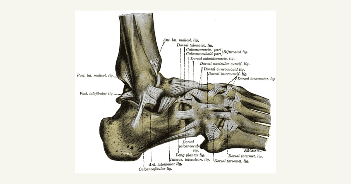
PTTD involves several structures in the foot and ankle, primarily the tibialis posterior tendon, which runs along the inner side of the ankle. Here’s a quick context of the foot and ankle anatomy.
The tibialis posterior tendon is just one component of a complex network of muscles, tendons, ligaments, and bones that work together to support movement and balance in the foot. Key structures involved include:
- Medial malleolus: The bony bump on the inside of the ankle, behind which the tibialis posterior tendon runs.
- Plantar fascia: A thick band of connective tissue that supports the arch of the foot.
- Spring ligament: A crucial ligament that helps maintain the arch of the foot alongside the posterior tibial tendon.
When PTTD occurs, the dysfunction weakens the support these structures provide, leading to flatfoot deformity and increased strain on other parts of the foot and ankle, including the lateral side of the ankle.
This anatomical interplay is vital for maintaining proper foot function and avoiding excessive stress on the musculoskeletal system during movement. When the tibialis posterior tendon fails to function correctly, it compromises the alignment and stability of the foot, causing pain, swelling, and difficulty with activities that require standing or walking.
Moreover, when this tendon degenerates, it can affect other structures like the flexor digitorum longus, flexor hallucis longus, the spring ligament, and the deltoid ligament, all of which contribute to the stability of the medial arch and overall foot function.
In some cases, soft tissues and bones such as the ankle bone and heel bone may also be impacted.
Stages of Posterior Tibial Tendon Dysfunction
As PTTD progresses, the severity of the tendon damage and associated deformities worsen, impacting both the foot’s structure and function. There are four stages of PTTD, each with different symptoms and treatment approaches.
Stage I
The posterior tibial tendon is inflamed, and pain is present, but the foot remains flexible with a normal arch. At this stage, the tendon is still functioning, but the inflamed tissue may cause discomfort during physical activity.
Pain relief and anti-inflammatory medication are a decision for your doctor or pharmacist. Custom arch support insoles may also be recommended to reduce strain on the tendon.
Stage II
The tendon starts to weaken and lose its ability to support the medial longitudinal arch. As a result, the arch begins to collapse, leading to a flexible flatfoot deformity. The affected foot may roll inward (ankle rolls). At this stage, the foot is still flexible, meaning that it can be realigned manually or with the help of orthotic devices like medial arch support insoles or braces.
Treatment focuses on preventing further progression of the deformity through nonsurgical interventions, such as a walking boot, custom orthotics, and physical therapy.
Stage III
The flatfoot becomes rigid, and tendon degeneration is more severe, with little to no flexibility. In this stage, the degeneration of the tendon is advanced, and secondary structures like the spring ligament and deltoid ligament may also be affected, leading to further instability of the foot and ankle.
Since nonsurgical treatments are often less effective at this stage, surgical intervention becomes a more likely option. Surgical reconstruction, such as tendon transfer or posterior tendon debridement, may be needed to improve function.
Stage IV
The deformity extends to the ankle joint, resulting in severe ankle deformities, pain, and limited ankle movements. The posterior tibial tendon is significantly damaged or nonfunctional, and the ankle joint may show signs of arthritis.
Surgical treatment is almost always required in Stage IV. Complex procedures like deltoid ligament repair, spring ligament reconstruction, or even ankle fusion may be necessary to rebuild stability and function.
Who Is More Likely To Develop PTTD?
PTTD is more common in adults, particularly those over 40, and women are more likely to develop the condition than men. This is due to factors such as hormonal differences, which can affect tendon elasticity and a higher prevalence of conditions like obesity and flat feet.
Those who have had previous surgery or injuries in the foot and ankle, such as Achilles tendon lengthening, are also at higher risk for PTTD.
Additionally, obesity, diabetes, joint disorders, steroid use, and inflammatory conditions like rheumatoid arthritis can also increase the chances of developing PTTD. For those with flatfoot deformity, foot and ankle specialists have noted that walking on uneven surfaces regularly may exacerbate the issue.
Causes of Posterior Tibial Tendon Dysfunction
The primary cause of PTTD is overuse of the posterior tibial tendon. This tendon wears and breaks down as we age from repetitive forces. Activities like running, walking, or standing for long periods, especially on uneven surfaces, can trigger and tear it more quickly.
Injury, tendon degeneration, or inflammatory conditions like posterior tibial tendonitis can also weaken the tibialis posterior tendon.
In some cases, an abnormal alignment of the foot, ankle joint, or heel bone can strain the tendon, leading to dysfunction.
Symptoms of Posterior Tibial Tendon Dysfunction
Foot and ankle pain is a broad symptom. For PTTD, the pain can feel more like the following:
- Pain along the inner side of the ankle or lower leg
- Swelling and tenderness in the affected foot
- A flattening of the medial arch
- Difficulty standing on tiptoes (single heel raise test)
- Difficulty climbing stairs
- Outward rolling of the ankle (ankle rolls)
- “Too many toes” sign, where more than the fifth toe is visible from behind
- Feeling of having uneven shoes
- A previous limp worsens
In later stages, the condition can cause severe pain, and the flatfoot deformity may become rigid and affect ankle movements. Further deterioration can develop arthritis in the foot or in severe cases, the ankles.
Diagnosing Posterior Tibial Tendon Dysfunction
Diagnosing PTTD requires a thorough examination by a foot and ankle specialist or surgeon. Early diagnosis is critical to prevent the condition from worsening. Below are tests your healthcare provider typically conducts to confirm the condition.
Tests for Diagnosing PTTD
Physical examination
The doctor begins by checking your foot for signs of swelling, tenderness, or pain along the posterior tibial tendon. They’ll also assess the flexibility of your foot and check for flattening of the arch, which is a common sign of PTTD. One common test is the single heel raise test. In this test, you’ll be asked to stand on one foot and lift your heel. Difficulty or inability to perform this test may indicate a weakened posterior tibial tendon.
Imaging tests
If the physical exam suggests PTTD, the doctor may order imaging tests to assess the extent of the damage. X-rays help reveal any changes in bone structure or flatfoot deformity. MRIs or ultrasounds are often used to get a detailed look at the condition of the posterior tibial tendon, showing signs of tendon degeneration or tears. They can also order a Computerised Tomography (CT) scan for 3D images of your soft tissues and bones, which is more detailed than X-rays.
Gait Analysis
The doctor may observe how you walk to spot abnormalities caused by a collapsing arch or uneven pressure distribution due to the weakened tendon. This can help assess how the condition affects your daily movements.
Questions To Ask During Consultation
To ensure you don’t miss out on details during a visit to your healthcare provider, the following questions can help you prepare:
- What stage of PTTD am I in?
- What is the root cause of my PTTD?
- Will I need surgery, or can nonsurgical treatments work?
- What activities should I avoid?
- How can I manage the pain?
- How long will recovery take with physical therapy?
- Are custom arch support insoles necessary for my condition?
Treatment for Posterior Tibial Tendon Dysfunction
Treating PTTD depends on its severity. In the early stages, nonsurgical options can effectively relieve symptoms, while more severe cases may require surgical intervention to correct the deformity and improve function. Let’s discuss these options below.
Non-Surgical Treatment
In the initial stages of PTTD, nonsurgical treatments often provide significant relief and help prevent further tendon damage. These approaches include:
- Rest and avoiding activities that put strain on the posterior tibial tendon, especially walking on uneven surfaces
- Immobilizing the foot with a walking boot or brace to reduce stress on the tendon and allow it to heal
- Stabilizing the foot with custom-made medial arch support insoles or orthotics
How Physiotherapy Helps
Physical therapy is a key component of PTTD treatment, particularly in the early stages. Phoenix Rehab in Singapore provides a tailored physical therapy program for tendon pain and injury recovery. Their methods involve a focus on targeted exercises for the tibialis posterior muscle, calf muscles, and flexor hallucis longus to improve tendon function and foot stability. These exercises not only enhance flexibility but also help prevent further degeneration by reinforcing the soft tissues surrounding the tendon.
Ankle sprain physiotherapy specialists may also guide patients in correcting gait abnormalities and reducing strain on the tendon during movement. They can also advise preventive measures and self-management techniques for tendon dysfunction.
Surgical Treatment
When non-surgical treatments no longer provide relief or the condition has progressed to a more advanced stage, surgery may be necessary. Several surgical options are available depending on the severity of the deformity and tendon damage:
What our patients say
Google reviews from our clinics in Singapore.


- Tendon transfer: In this procedure, foot and ankle surgeons replace the damaged posterior tibial tendon with a healthy tendon, often the flexor digitorum longus, to improve functionality and support the arch.
- Posterior tendon debridement: Surgeons remove damaged or inflamed tissue from the posterior tibial tendon to promote healing and improve tendon strength.
- Spring ligament reconstruction or deltoid ligament repair: These surgeries focus on repairing the ligaments that have been affected by the progressive collapsing foot deformity, helping rebuild stability to the ankle and foot.
- Medial displacement calcaneal osteotomy: This procedure realigns the heel bone to correct the flatfoot deformity, providing better structural support for the foot.
Recovery Period for PTTD
Recovery times vary depending on the treatment approach. Nonsurgical treatments, like physical therapy and orthotics, may take several months to show improvement. Very mild cases can go on for 6-8 weeks.
Surgical procedures, on the other hand, require longer recovery periods. Patients may need to wear a short leg cast or walking boot for several weeks post-surgery, followed by physical therapy to improve mobility and strength.
Full recovery can take anywhere from six months to a year, depending on the complexity of the surgery and the patient’s overall health.
How Can You Prevent Posterior Tibial Tendon Dysfunction?
Preventing PTTD involves maintaining a healthy lifestyle and taking care of your feet. Use proper footwear with medial arch support, especially if you have flat feet. Avoid walking on uneven surfaces for extended periods, and practice exercises to strengthen the tibialis posterior muscle and calf muscles.
If you notice any pain or swelling in the ankle or foot, consult an ankle specialist early to prevent the condition from progressing.
Conclusion
PTTD can be a progressive disorder if left untreated. Early diagnosis and appropriate treatment can prevent further complications and help you maintain healthy foot function. Whether through nonsurgical treatments or surgical reconstruction, managing PTTD with the guidance of a foot and ankle surgeon and physical therapists can help you throughout the journey of improving your mobility.
Where are you with this right now?
1. It is settling on its own
Most injuries and most flare-ups settle over weeks with sensible load management and a return to normal movement. Improvement is judged over a month, not over a day, and good days and bad days inside that are usual. If you are doing more this month than last, and sleeping better, it is behaving the way it should. You do not need us for this, and we would rather say so.
2. It has stopped improving
You have adapted without noticing. The other side takes the load, you plan movements you used to make without thinking, and you have quietly dropped things you used to do.
Pain is the first thing to leave. Movement and strength are the last things trained back, and most people stop before that part is done. That is the gap worth measuring, and it is not measured by how you feel on the day.
The Stalled Recovery Review is three appointments over three weeks with the same therapist, because what a joint does under load cannot be read in one visit. The first finds what is limiting it. The next two measure how it responds. $690, nett, no GST, which is three appointments at $230.
3. You have had surgery, or you have a date
Recovery after an operation has a shape, and knowing which stage you are in tells you whether you are on it. See the recovery programs, hand and physiotherapy, or the four stages and what each one works on if you are not sure which applies to you.
All of it runs on the same four stages. The Phoenix Recovery Protocol explains what each stage works on and what you have to meet to move up. No referral needed to start.
Next step
Find out which stage you are in
Pain is the first thing to leave. Movement and strength are the last things trained back, and most people stop before that part is done. One Assessment and Treatment is a single 45-minute appointment at $230, nett, no GST. Your therapist finds what is actually limiting you, treats it in the same appointment, and you leave with your current stage and a written plan.
Prefer to phone? Each of our five clinics has its own number on the clinics page. No referral needed to book.
Related reading
Where we are
Clinics across Singapore
ORCHARD
400 Orchard Road #12-12
Orchard Towers, S238875
NS22 Orchard MRT
Physiotherapy and hand therapy in Orchard →
WhatsApp 8800 1830TAMPINES
9 Tampines Grande #01-20
Asia Green, S528735
EW2DT32 Tampines MRT
Physiotherapy and hand therapy in Tampines →
WhatsApp 8780 9608SERANGOON
265 Serangoon Central Drive #04-269
S550265 · Lift C only
NE12CC13 Serangoon MRT
Physiotherapy and hand therapy in Serangoon →
WhatsApp 8892 4121TANJONG PAGAR
10 Anson Road #02-77A
International Plaza, S079903
EW15 Tanjong Pagar MRT
Physiotherapy and hand therapy in Tanjong Pagar →
WhatsApp 8227 7074JURONG EAST
2 Venture Drive #02-02
Vision Exchange, S608526
NS1EW24 Jurong East MRT
Opening 25 October 2026
Physiotherapy and hand therapy in Jurong East →
WhatsApp 8242 7340