Humerus Fracture Physiotherapy
By Nigel ChuaA humerus (or humeral) fracture refers to a break in the bone of the upper arm. The humerus bone is the long bone that is located between the shoulder and the elbow.
Humeral fractures can be categorized into three type of groups, depending on which part of the bone is broken.
A proximal humerus fracture is when the humerus bone is broken at or near the shoulder. Mid-shaft humeral fractures, also known as humeral shaft fractures or diaphyseal humerus fractures, refer to a break in the middle section of the bone. Distal humerus fractures occur when the humerus bone is broken near to the elbow.
About 3 - 5% of all broken bones are mid-shaft humeral fractures, and these types of fractures tend to affect two groups of people:
- young males (20-30s) who have experienced a high-energy or high-impact type of injury
- elderly females (60-70s) with low-energy or low-impact types of injuries.
HUMERAL SHAFT ANATOMY AND REVIEW
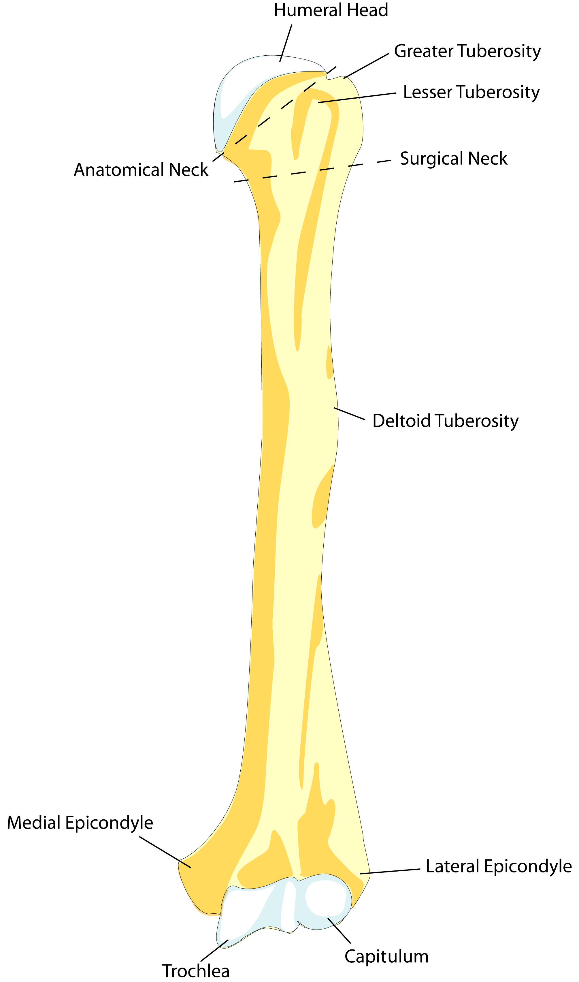
The mid-shaft region of the humerus is the long, thin part of the bone.
The top part is cylindrical in shape, and as it goes downwards towards the elbow, it becomes narrower and more prism-shaped. The back surface of the shaft of the humerus is bigger than the front part of the bone.
The shaft of the humerus can be divided into thirds:
- the proximal (upper) third
- the middle (mid) third
- the distal (lower) third.
COMMON CAUSES OF MID-SHAFT HUMERUS FRACTURES
Mid-shaft humerus fractures are usually the result of:
- Falls: The most common cause of a mid-shaft humeral fracture, particularly in the elderly, is a simple fall. Occasionally it may be caused by falling onto an outstretched hand when the arm is abducted (out to the side).
- Direct Forces and Blows: High-energy forces, such as in sports injuries and motor vehicle accidents that affect the upper arm, often cause transverse fractures.
- Torsion (Rotational) Forces: Forceful twisting of the upper arm can lead to a long spiral fracture of the humerus. Long spiral humeral fractures in children should be carefully examined, as they may indicate child abuse, neglect and social issues.
- Bone Disease: Metastatic breast cancer can cause spontaneous fractures in the middle of the humerus.
CATEGORIZATION OF HUMERUS FRACTURES
Mid-shaft humeral fractures can be classified into different types depending on the location and direction of the break and the associated damage:
1. Location
- Proximal Third: 30% of humeral fractures occur in the top third region
- Middle Third: 60% of mid-shaft humerus fractures occur in the middle portion
- Distal Third: 10% of humeral shaft fractures occur in the lower third.
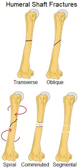
2. Fracture Pattern
- Transverse Fracture: where the line of the fracture runs perpendicular to the bone shaft i.e. horizontally across the humerus
- Oblique Fracture: where the fracture line is angled
- Spiral Fracture: where there is a spiral-shaped fracture line - it seems to "go around the bone"
- Comminuted Fracture: where the bone breaks into several pieces/fragments
- Segmental Fracture: where there are at least two fracture lines that together isolate a section of bone
3. Degree of Displacement
A displaced humeral fracture happens when the fractured humeral bone fragments of the shaft do not line up normally. A non-displaced fracture is where normal alignment is maintained despite the fracture line.
4. Soft Tissue Damage
- Open Fractures: Open fractures happen when the broken bone punctures through the skin from the inside. They can also be caused by a blow to the shoulder that cuts through the skin and muscle. Around 3-10% of humeral shaft fractures are open fractures.
- Closed Fractures: Most humeral fractures are closed fractures, where the skin remains unbroken.
5. Pathological Fracture
This type of fracture occurs when the bone has broken due to a disease that has weakened the bone, such as metastases and cancer. The fracture may occur spontaneously, so there may not necessarily be a specific incident that has directly caused the injury.
SYMPTOMS OF MID-SHAFT HUMERAL FRACTURES
Whichever part of the bone is broken, a mid-shaft humerus fracture will typically cause:
- Pain: There is usually instantaneous, severe, ongoing pain associated with the fracture. Patients will know that they have hurt themselves badly, and have potentially fractured their bone.
- Restricted Arm Movement: Shoulder and arm movement will result in a lot of pain, so typically patients with a mid-shaft humerus fracture will be protective and often unwilling to move their arm. There will be associated weakness throughout the whole arm including the wrist and hand.
- Swelling And Bruising: There will be significant swelling and bruising in the upper arm which may travel and go all the way down to the hand. The swelling usually comes on fairly quickly, and this is due to extensive soft tissue damage and bleeding in the surrounding area.
- Bony Sounds and Sensations (Crepitus): Patients may experience a grinding, grating sound and sensation when the fractured pieces rub against each other directly.
- Obvious Fracture Deformity: If the mid-shaft humeral fracture is displaced (moved), there may be an obvious deformity of the upper arm or it may look abnormal e.g. the arm may appear shorter than normal if the bone at the fracture site has overlapped.
- Bleeding: If the humeral fracture is an open fracture where the bone has pierced through the skin (or if the blow or force has split the skin and soft tissue), there will be bleeding.
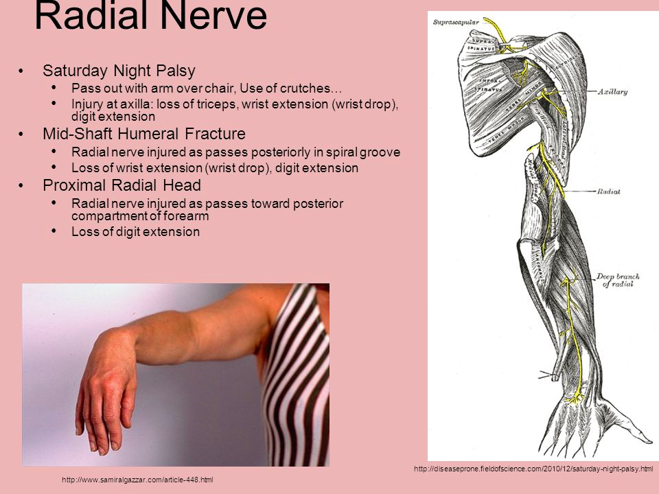
- Altered Sensation: If there is nerve damage associated with the humerus fracture, there may be altered sensation such as pins and needles or numbness at or below the fracture site. Normal movement at the wrist and hand may also be affected as the motor aspect of the nerve can be injured. Unfortunately, radial nerve damage is fairly common with humeral shaft fractures.
DIAGNOSING HUMERAL SHAFT FRACTURES
Sometimes mid-shaft humeral fractures can be obvious, especially when there is an obvious fracture deformity. However in many cases, the attending doctor will need to confirm these fractures with imaging.
If you doctor suspects a mid-shaft humerus fracture, you will be sent for x-rays.
X-rays will be taken in different directions, usually from front to back (AP) and from the side (lateral). Both the shoulder and elbow joints should also be evaluated for any damage.
X-rays help the doctor to see where the fracture is, what type of fracture it is, any associated damage and the severity of the injury so that they can plan the best course of treatment.
COMMON TREATMENTS OF MID-SHAFT HUMERUS FRACTURES
Treatment for humerus fractures will vary a little depending on the location and severity of the fracture, but the good news is that in most cases, surgery is not required. Approximately 90% of humeral shaft fractures unite (heal) without the need for surgery, and just with shoulder humeral physiotherapy.
Non-Surgical Treatment
Non-surgical treatment for a mid-shaft humerus fracture usually consists of:
1) Immobilisation
For the first 4 weeks, patients will need to protect and immobilize the humeral mid-shaft fracture to allow the fracture to start to form and swelling and pain to subside.
Humeral shaft fractures are typically treated with a coaptation splint that extends from the shoulder to the forearm, and holds the elbow in 90 degrees flexion.
In some cases, you may also be given a collar and cuff sling to support the arm.
It is very important to let the arm hang by your side without any support through the elbow, as this allows gravity to help realign the fracture. Often patients try to "support" the fracture by tightening their biceps and/or elbow, but that is not the correct way to encourage humeral fracture healing.
If there is any pressure through the elbow it pushes the bones together, resulting in the bones healing in the wrong position.
After two to three weeks, the coaptation is switched for a functional brace or a cylindrical brace that fits around the upper arm, holding the humerus in place. It leaves the shoulder and elbow free to move, which helps to prevent stiffness.
2) Medication
You will be prescribed painkillers and anti-inflammatories. If you have an open fracture you will be given additional antibiotics to reduce the risk of infection.
3) Exercises
Our senior physiotherapists in Phoenix Rehab will work together with you through a shoulder physiotherapy program after your mid-shaft humeral fracture.
We will help you get started with active exercises for the elbow, wrist and hand to prevent stiffness and weakness from developing, and it is usually fine to take your arm out of your sling to do these exercises.
Once your orthopedic doctor had determined that your humeral fracture has started to unite, they will let us know and we will get you started on shoulder mobility exercises. We usually start shoulder exercises with very safe pendulum exercises where patients use gravity to move the arm, and active assisted exercises where you use your good arm to support and move the broken arm.
After just a few weeks, we will be able to progress you with progressive strengthening exercises and more advanced range of motion exercises.
It is very important to stick with the shoulder physiotherapy program and do your shoulder exercises every day, until you have regained full strength, flexibility and movement of the shoulder, elbow and hand.
If you stop too soon, or only do your exercises sporadically, you are likely to have ongoing limitations in the mobility, strength and function of your arm.
Surgical Treatment
Around 10% of humeral shaft fractures will require surgical treatment.
If the bone fragments have moved or are displaced, the broken fragments will need to be realigned and held in place. This process is typically known as an "ORIF", which stands for open reduction internal fixation.
Most of the time, the humeral fracture is secured with either:
Metal Plates
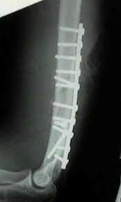
Humeral shaft fractures that require surgery are usually treated with a large metal plate that is securely held in place by screws.
- Pros: Highest success rate for surgical treatment of humerus fractures.
- Cons: Higher risk of nerve damage and non-union than non-surgical treatment.
Intramedullary (IM) Rods
In some cases a humerus fracture will be treated surgically with an intramedullary rod/nail. This is when a long metal rod is placed down the middle of the bone. IM nails can be used to stabilise a humerus fracture that is between 2cm below the surgical neck and 3 cm above the elbow.
- Pros: Less invasive and less chance of nerve damage.
- Cons: Lower healing rate and higher rate of non-union.
These implants are designed to hold the bones together while the fractured parts heal, and patients should expect full union to be achieved.
Metal implants are not intended to be a long term solution, and they tend to need to be removed. If the bones fail to unite, there is a high chance that the implant will at some point fail and further surgery may be required.
The usual indications for treating a mid-shaft humerus fracture surgically are:
- If the Humeral Fracture Can’t be Reduced / Aligned: In some cases, it may be difficult to realign the bones, or maintain the realigned position. This is usually due to associated soft tissue injuries, head injuries, secondary injuries or patient obesity.
- Open Fractures and Bleeding: If the bone has shifted enough to pierce and puncture the skin, it is unlikely that the bone will realign without surgery, and it is likely severe enough to warrant immediate corrective surgery.
- Segmental Fractures: If the fracture has caused a fragment of bone to break off, it will often need to be secured surgically.
- Multiple Fractures: If the humerus has broken into four or more pieces, if both the left and right upper arms are broken, or there is a break in one of the forearm bones on the same side, then surgery is usually necessary.
- Vascular Damage: If there is significant vascular damage, surgery will be required.
- Nerve Damage: If the brachial plexus has been damaged, surgery is usually necessary. If the radial nerve has been damaged, it does not usually require surgical intervention unless there are associated injuries – the radial nerve damage will most likely heal within 3-4 months without the need for surgery.
- Non-Union: If after a few weeks the humeral shaft fracture has failed to heal and the bones haven’t united, surgery may be necessary to stabilise the bone.
- Skin or Soft Tissue Damage: If the skin or soft tissues of the upper arm have been damaged to the extent that it is not possible to wear a splint, e.g. severe burns, surgery may be required.
RECOVERY PROCESS
Over 90% of humerus fractures treated non-operatively will unite and are usually fully healed (complete union) within 8-12 weeks. Though older patients may not regain full 100% shoulder movement, they usually regain enough functional range for their day to day activities (we will always aim for 100% of course).
Complications are more common with complex fractures and those requiring surgery. Orthopedic surgeons will tend to be more conservative and try to prevent unnecessary surgeries.
Mid-shaft fractures may heal with slight angulation. Although healing may not be completely straight, this doesn’t usually cause any functional issues as the shoulder and elbow accommodate.
COMPLICATIONS OF MID-SHAFT FRACTURES
At the Time of Injury
Nerve Damage
There is often associated damage to the surrounding nerves with mid-shaft humerus fractures.
The most commonly damaged nerve is the radial nerve, as the nerve wraps around the back of the humerus, with between 8-15% of mid-shaft fractures resulting in radial nerve damage.
Injury to the radial nerve usually occurs at the time of injury, but can also occur when the fracture is reduced, so great care should be taken when realigning the bones.
Blood Vessel Damage
There may be damage (or risk of damage) to the brachial artery.
During Recovery
- Frozen Shoulder: This occurs when the capsule of the shoulder joint becomes inflamed and thickened, typically due to lack of shoulder movement after a shoulder fracture.
- Myositis Ossificans: This occurs when calcification develops in the surrounding muscles. Myositis ossificans is usually caused by returning to activities too quickly after a mid-shaft humerus fracture.
- Fracture Angulation: Often the bone heals at a slight angle, but it rarely has any functional impact and is usually not visibly noticeable.
- Mal-Union or Non-Union: Mal-union is when the bone fragments unite significantly out of normal alignment. Non-union is where the bone fragments fail to unite back together. Non-union is rare with a mid-shaft humerus fracture, occurring in only 4% of cases. It tends to occur with a proximal third mid-shaft fracture or spiral humerus fracture.
SHOULDER HUMERAL FRACTURE PHYSIOTHERAPY
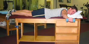
Patients may also receive the following physiotherapy treatment modalities:
- cold therapy
- moist heat paraffin wax therapy
- radio-frequency Indiba physiotherapy to accelerate soft tissue healing
- joint mobilization
- stretching exercises
- strengthening exercises
- scar management
- hands-on manipulation and mobilization (manual therapy)
- computerized spinal decompression traction
- soft tissue management
- heat therapy, heat treatment and heat packs to relieve tight muscles and joints
- ultrasound therapy to accelerate soft tissue healing
- exercise therapy
- acupuncture and/or dry needling
- deep tissue release
Browse other articles by category
Physiotherapy for Knee Pain Physiotherapy For Slipped Disc Physiotherapy for Neck Pain PHYSIOTHERAPY
 Hand Therapy
 Alternative
 Massage
 Traditional Chinese Medicine Treatment
 Rehab
Rehab
 Physiotherapy For Lower Back Pain
Physiotherapy For Shoulder Pain
Orthopedic Doctors, Insurance & Healthcare
Physiotherapy For Upper Back Pain
Frozen Shoulder
Physiotherapy for Back Pain

 Whatsapp us now